|
During dialysis rounds, 64-year-old Mrs. D complained of a painful sore on her left breast. Mrs. D was a thin, white woman who had been on hemodialysis for four years. She noted not only the tenderness and extreme pain in her breast, but also complained that this was accompanied by a significant amount of pain in her thighs.
Mrs. D denied any recent trauma, reported no new exercise routines, or changes in medication. A review of her chart showed an episode of atrial fibrillationtwo months ago that was treated with warfarin therapy, but otherwise her medical history for the past year was unremarkable.?
1. Examination and Laboratory Tests?
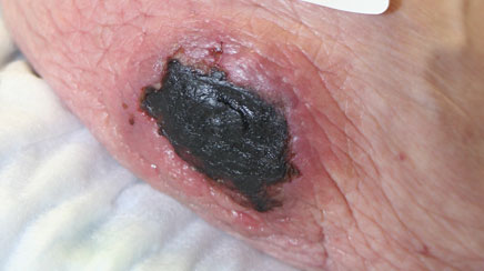
Examination of the left breast showed a black area with a red circumference without any drainage (Figure 1). The breast area was exquisitely tender. There were no palpable lymph nodes, and the right breast was slightly lumpy without any open lesions.
Bilateral thighs had multiple moveable lumps, which were firm and approximately ?3X4 cm each. The thigh masses were also hypersensitive to touch, but without accompanying skin lesions.
Mrs. D's lungs were clear to auscultation. Her heart rate was regular without any murmur or ectopy. She dialyzed through an upper left arm arteriovenous fistula. The rest of the examination was noncontributory.
Mrs. D's hemoglobin level was 11.5 g/dL. (In adult women, the normal range is 12-16 g/dL.) Her calcium level was below normal at 7.8 mg/dL (baseline range 8.4-10.2 mg/dL), and her phosphorus level was significantly elevated at 7.6 mg/dL (3.5-?5.5 mg/dL is the typical target range for dialysis patients).
Mrs. D's intact parathyroid hormone (PTH) level was 1251 pg/ml (normal is 14-?72 pg/ml), her albumin level was 2.5 g/dL (normal 3.5-5.2 g/dL) and her hemoglobin A1c was 5.9% (standard range of 4.8%-5.9%). ?
2. Differential Diagnosis?
The differential diagnosis includes cholesterol emboli, necrosis from warfarin, cellulitis, nephrogenic sclerosing fibrosis (NSF), vasculitis, calciphylaxis or gangrene.1
Cholesterol emboli and their close relatives (atherosclerosis and atheroemboli) can be excluded due to location of the lesion on the upper extremity, as well as intact peripheral pulses. Gangrene is also excluded for this reason.
Vasculitis is unlikely since the patient lacks the tell tale signs — neuropathy and polyartertis — plus Mrs. D is older than the usual age group. NSF is also unlikely due to a lack of recent radiology tests. Heparin-induced necrosis was considered, but ?Mrs. D's platelet count came back in the normal range, which made this diagnosis less likely. Cellulitis and calciphylaxis are usually hard to distinguish from each other without a biopsy, but the lumpy lesions in the patient's thighs made calciphylaxis the most likely diagnosis. ?
3. Treatment and Outcome?
Calciphylaxis is an ischemic disease of the arterioles. Calcification of the vessels causes narrowing, decreased blood flow and hypoxia, leading to necrosis of the skin. Since calciphylaxis has an 80% death rate once ulceration occurs, all known treatments were initiated simultaneously.2 Intravenous (IV) sodium thiosulfate was started (12.5 mg with the first dialysis, then increasing to 25 mg with each subsequent hemodialysis).
Mrs. D was referred to the local wound center and a breast surgeon. It is unknown how sodium thiosulfate works in calciphylaxis, but it is thought to dissolve the calcification in the tissues and move the previously insoluble calcium into the bloodstream.3 Empiric reports of the effectiveness of sodium thiosulfate treatment were a clinical guide here, although no rigorous, controlled studies have been conducted.
Mrs. D's warfarin was discontinued since it is a known factor in the development of calciphylaxis. Mrs. D was started on oral cinacalcet (Sensipar), a calcimimetic that increases the sensitivity of the parathyroid gland to calcium.4,5 However, she was unable to tolerate cinacalcet, so it was stopped.
Pain control was an extremely important and difficult problem in our patient due to severe nausea and vomiting, thus a palliative care consult was obtained. A combination of patches and oral narcotics, both short and long acting, were needed for pain management. ?
The wound center began hyperbaric oxygen (HBO) treatments and local care of the lesions. There are case reports that indicate that HBO helps wound healing by increasing oxygenation to the tissues affected by calciphylaxis.6 The breast surgeon was reluctant to operate due to the likelihood of poor wound closure and healing. He asked that we consider a parathyroidectomy prior to any breast surgery, unless the wound center was able to heal the lesions with conservative care.?
Mrs. D's breast lesions went from bad to worse over a two-year period. The open, necrotic tissue on the left breast gradually encased the entire breast and small lesions started forming on the right breast (Figure 2).

Slowly, both breasts turned black from necrosis. The patient was in incredible pain with the lesions now manifesting as hard sores. Immediately after the IV sodium thiosulfate was started, the lumps on Mrs. D's breasts and thighs started to decrease and her leg pain diminished. ?
With the obvious failure of the conservative care (discontinuation of warfarin, HBO, local wound care, sodium thiosulfate, and attempted use of cinacalcet), it was time for a more aggressive approach. A parathyroidectomy was planned, but the anesthesiologist on call was reluctant to put Mrs. D under general anesthesia. However, when weighing the high-risk of death from calciphylaxis versus the anesthesia risks, the lesser of two evils was chosen. ?
Mrs. D's parathyroid gland was removed and she experienced a slightly rocky postoperative course. On the positive side, the accumulated calcium, which had painfully hardened the patient's breast tissue, markedly decreased within the first 24 hours. Mrs. D's intact PTH level dropped after surgery from 1251 pg/ml to 231 pg/ml (dialysis dependent patient goal 150-600 pg/ml).7 After the parathyroidectomy, the 'hungry bone' syndrome that occurs caused the calcium in the tissues to reabsorb, and necrosis in the patient's breast tissue was thwarted.
HBO was restarted and Mrs. D's breast tissue developed a demarcation line between the dead necrotic tissue and the tissue that had healed. After eight days, the breast surgeon brought Mrs. D back to the operating room for a bilateral mastectomy. As he commented to Mrs. D after surgery, "Your breasts were so hard, I could have used them as bowling balls." This comment clearly validates the characterization of this disease with the phrase, "When man turns to stone."8?
4. Discussion?
Although calciphylaxis is a dermatological diagnosis, most dermatology practitioners have never seen a case. Yet, most nephrology practitioners can identify calciphylaxis simply by viewing a lesion.
Calciphylaxis is seen in 1%-4% of long-term dialysis patients, but can also be seen in patients with autoimmune diseases such as Crohn's disease or lupus nephritis, cancer, hypercoagulable states and in parathyroid disease.9 Traditional risk factors for calciphylaxis, besides uremia, are as follows: female sex, obesity, Caucasian race, high phosphorus and/or intact PTH level, maintaining on medications such as warfarin, vitamin D analogues, oral calcium and systemic corticosteroids.10 ?
The natural course of the disease is indolent with a one-year survival rate of less than 50%, even with treatment.11 The pain that accompanies the lesion(s) cannot be overstressed. It is searing and continuous, as though a part of the body is clotted off.
Treatment is multi-factorial and various specialists need to be involved to maximize the chance of wound healing. With the high rate of death and limb loss associated with calciphylaxis, the more aggressive the treatment, the better the chance of overall and limb survival.12?
Prevention is vital, since survival rates are poor. While calciphylaxis is more common among dialysis patients, cases that occur in patients without renal disease are more likely to be missed. A report on breast calciphylaxis that occurred after coronary artery bypass graft was recently published.13 At present, there are no specialized laboratory tests for diagnosing calciphylaxis; the patient's phosphorus, calcium and/or intact PTH levels may be elevated, but just as often, they are in the normal- or low-range.14
Skin necrosis, typically referred to as metastatic calcification can occur at normal levels.15 Even with normal readings, calcium and phosphate crystals can progressively accumulate in the small blood vessels of the fat tissue and skin. A high index of suspicion must be entertained when confronted with a suspicious lesion.?
5. Summary?
Mrs. D has been out of treatment for two years now. She spent two months in rehabilitation after her episode of calciphylaxis and was then weaned off of pain medications. She claims to be the only patient ever who was thrilled to have a bilateral mastectomy. She is now teaching phosphorus control to the other dialysis patients at the unit, and often states that she counts her blessings that she was one of the few who survived. ?
Kim Zuber, MS, PA-C, and Jane Davis, DNP,are nephrology practitioners and Executive Council members of the National Kidney Foundation in Alexandria, Va. Bill Bartow, PA-C, is the Clinical Director of the INOVA Wound Healing Center in Annandale, Va.?
References?
1. Fisher AH, Morris DJ. "Pathogenesis of calciphylaxis: study of three cases with literature review." Hum Pathol. 1995;26:1055-1064.?
2. Ross EA. "Evolution of treatment strategies for calciphylaxis." Am J Nephrol. 2011;34:460-467.
3. Singh RP, Derendorf H, Ross EA. "Simulation-based sodium thiosulfate dosing strategies for the treatment of calciphylaxis." Clin J Am Soc Nephrol.2011;6:1155-1159.?
4. Raymond CB, Wazny LD. "Sodium thiosulfate, bisphosphonates and cinacalcet for treatment of calciphylaxis." Am J Health Syst Pharm. 2008;65:1419-1429.?
5. Banerjee C, Woller SC, Holm JR et al. "Atypical calciphylaxis in a ?patient receiving warfarin then resolving with cessation of warfarin ?and application of hyperbaric oxygen therapy." Clin Appl Thromb Hemost. 2010;16:345-350. ?
6. Podymow T, Wherrett C, Burns KD. "Hyperbaric oxygen in the treatment of calciphylaxis: a case series." Nephrol Dial Transplant. 2001;16:2176-2180.
7. National Kidney Foundation. "NKF, K/DOQI clinical practice guidelines for chronic kidney disease: evaluation, classification, and stratification." ?Am J Kidney Dis. 2002;39:S1-S266.?
8. Floege J. "When man turns to stone: extraosseous calcification in uremic patients." Kidney Int. 2004;65:2447-2462.?
9. Nigwekar SU, Wolf M, Sterns RH et al. "Calciphylaxis from nonuremic causes: a systematic review." Clin J Am Soc Nephrol. 2008;3:1139-1143.
10. Weenig RH, Sewell LD, Davis MD, et al. "Calciphylaxis: natural history, risk factor analysis, and outcome." J Am Acad Dermatol. 2007;56:569.?
11. Sewell LD, Davis MD, McCarthy JT, Pittelkow MR. "Calciphylaxis: ?natural history, risk factor analysis and outcome." J Am Acad Dermatol. 2007;56:569-579.?
12. Goel SK, Bellovich K, McCullough PA. "Treatment of severe metastatic ?calcification and calciphylaxis in dialysis patients." Int J Nephrol.2011;24:701603.
13. Cathenis K, Goossens D, Vertriest R, et al. "Breast infarction due to calciphylaxis after coronary artery bypass grafting." Ann Thorac Surg. 2011;91:1603-1606.?
14. Kalajian AH, Malhotra PS, Callen JP et al. "Calciphylaxis with ?normal renal and parathyroid function: not as rare as previously believed." Arch Dermatol. 2009;145:451-458.
15. Fine A, Zacharias J. "Calciphylaxis is usually non-ulcerating: risk ?factors, outcome and therapy." Kidney Int. 2002;61:2210-2217.
?All electronic document accessed May 15, 2012.?
|
 Log in to explore the world's most comprehensive database of dialysis centres for free!
